Sponsored by
Pseudo Color
“As an artist, working in a medical facility seeing both the eye’s fragility and its resilience provides a sense of the fleeting and the profound. The anatomical structures of the human eye are intriguing to me not only in their function but also their form. With artistic license (and a removal from context) these photographs become abstract, yet intimately familiar explorations of color and form.” Kelly Aileen Oldstein, Certified Ophthalmic Photographer at Chester County Eye Care and Owner, Kelly Aileen Photography, West Chester, PA, USA.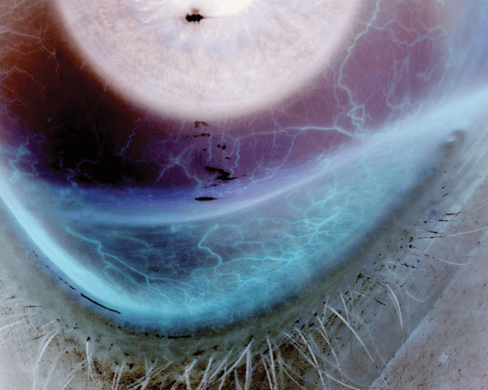
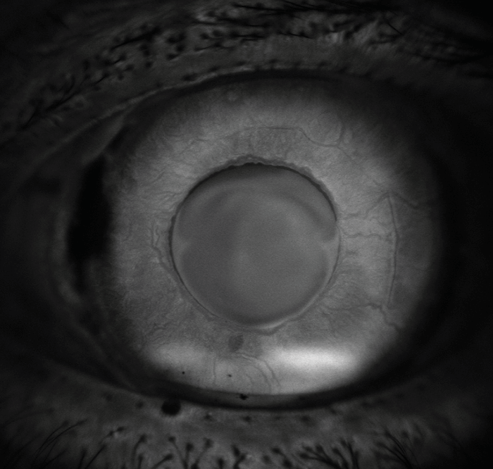
So Close and Yet So Far
One look at the displaced IOL and glaucoma implant shows that this eye might have seen an ophthalmologist or two. Houston Sharpe III, Ophthalmic Imaging Specialist, UNC Kittner Eye Center, Chapel Hill, NC, USA.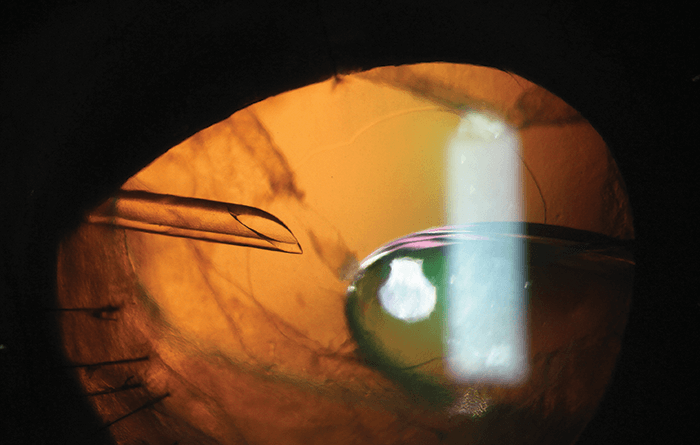
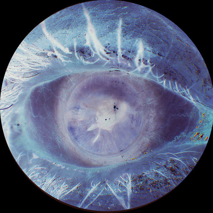
Swimming in a Blue Sea
Dendritic ulcer and unstable tear film after instillation of fluorescein. Helena Prior Filipe, Department of Ophthalmology, Hospital of the Armed Forces, Lisbon, Portugal.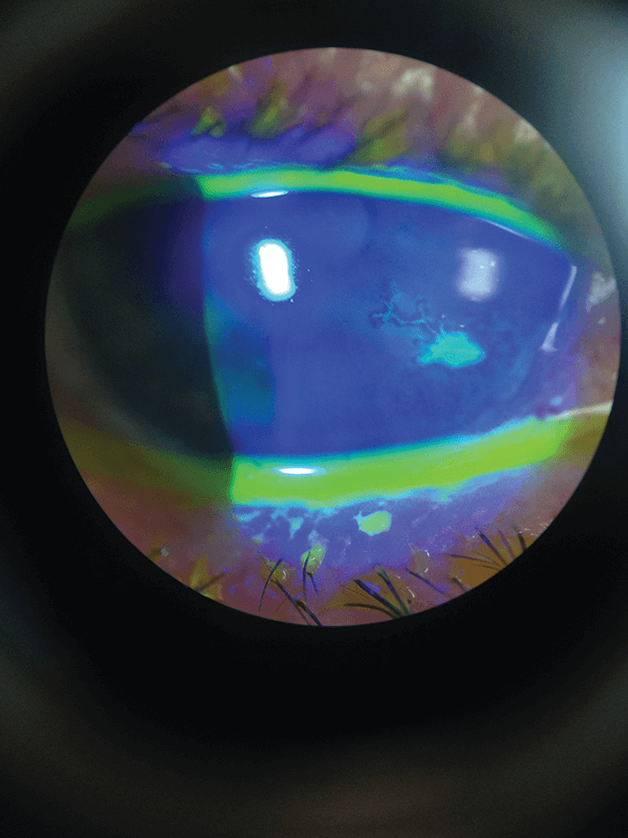
Rubeosis Iridis
Fluorescein angiography of neovascularization of the iris. Zachary Dupureur, Michael Tsipursky and Mary Bruce, Carle Foundation Hospital, Urbana, Illinois, USA.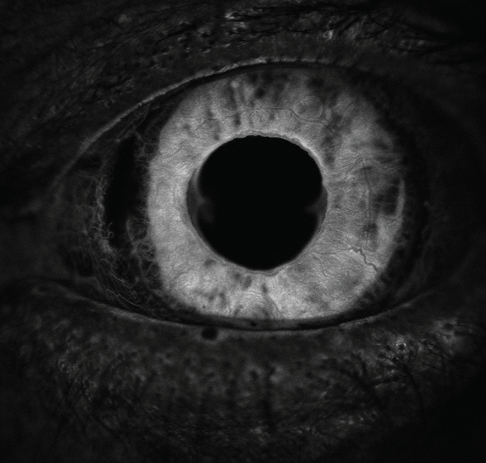
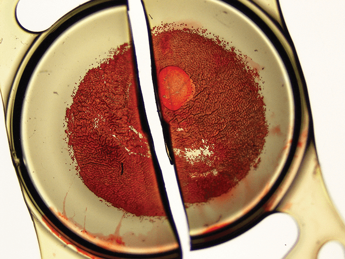
Hyphema
This patient presented with hyphema caused by a rock striking the eye that was propelled by a weed whacker. Kelly Aileen Oldstein, Certified Ophthalmic Photographer at Chester County Eye Care and Owner, Kelly Aileen Photography, West Chester, PA, USA.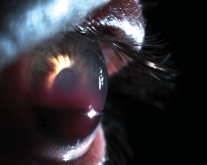
Beauty in the Breakdown
An inverted-color image of a patient with an iris defect and corneal scarring creates an image ofimmense beauty. Kelly Aileen Oldstein, Certified Ophthalmic Photographer at Chester County Eye Care and Owner, Kelly Aileen Photography, West Chester, PA, USA.
Well, There’s Your Problem
Viewing this slit beam from the side gives a much better appreciation of the elevation of the cornea. Houston Sharpe III, Ophthalmic Imaging Specialist, UNC Kittner Eye Center, Chapel Hill, NC, USA.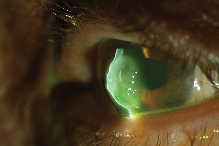
Can You See Me Now?
Using bright diffuse lighting, a clear visualization is captured of this one day postoperative AC-IOL. Houston Sharpe III, Ophthalmic Imaging Specialist, UNC Kittner Eye Center, Chapel Hill, NC, USA.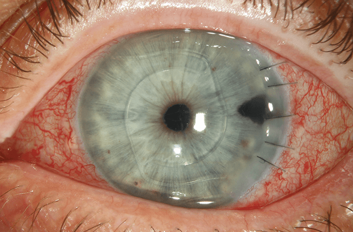
Localized Calcification of a Hydrophilic Acrylic IOL
Some months after uneventful implantation of this hydrophilic acrylic IOL, the patient underwent further surgery for a retinal detachment repair, including vitrectomy with silicone oil tamponade. A few months later, the IOL exhibited a localized, central, round area of opacification on the anterior surface. Due to decreased visual function, the lens was explanted, and staining with alizarin red demonstrated that calcification caused the opacification. IOL explanted by Robert Cionni and analyzed by Liliana Werner and Nick Mamalis, Salt Lake City, UT, USA.