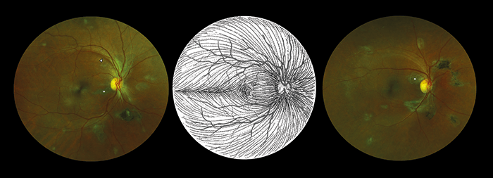
What and why? Case-control prospective study examining whether any specific retinal signs can be attributed to past Ebola virus disease in survivors and whether the virus persists in the aqueous humor (1). Who? Eighty-two Ebola virus disease survivors with post-Ebola syndrome who had previously reported ocular symptoms, and 105 unaffected and asymptomatic controls. How? Ocular examinations, including widefield retina imaging (scanning laser ophthalmoscopy) and OCT analysis; paracentesis of the anterior chamber was performed on two patients with white cataract.
Findings? A novel retinal lesion was identified in 14.6 percent of Ebola virus disease survivors; the scarring followed the pattern of optic nerve axons (Figure 1). The aqueous humor sampled from two Ebola virus survivors with white cataract was negative for viral RNA.
Upshot? According to corresponding author Paul Steptoe (2), “The distribution of these retinal scars or lesions provides the first observational evidence that the virus enters the eye via the optic nerve to reach the retina in a similar way to West Nile Virus. Luckily, they appear to spare the central part of the eye so vision is preserved.” He added, “Our study also provides preliminary evidence that in survivors with cataracts, aqueous fluid does not contain Ebola virus, therefore enabling access to surgery.”
References
- PJ Steptoe et al., “Novel retinal lesion in Ebola survivors, Sierra Leone”, Emerg Infect Dis, (2017). DOI: 10.3201/eid2307.161608. University of Liverpool, “Ebola survivors have a ‘unique’ retinal scar”, (2017). Available at: http://bit.ly/EbolaScar. Accessed May 17, 2017.
