Conexiant
Login
  • Corneal Physician
  • Glaucoma Physician
  • New Retinal Physician
  • Ophthalmology Management
  • Ophthalmic Professional
  • Presbyopia Physician
  • Retinal Physician
The Ophthalmologist
  • Explore

    Explore

    • Latest
    • Insights
    • Case Studies
    • Opinion & Personal Narratives
    • Research & Innovations
    • Product Profiles

    Featured Topics

    • Anterior Segment
    • Glaucoma
    • Retina

    Issues

    • Latest Issue
    • Archive
  • Subspecialties
    • Cataract
    • Cornea
    • Glaucoma
    • Neuro-ophthalmology
    • Oculoplastics
    • Pediatric
    • Retina
  • Business

    Business & Profession

    • Professional Development
    • Business and Entrepreneurship
    • Practice Management
    • Health Economics & Policy
  • Training & Education

    Career Development

    • Professional Development
    • Career Pathways

    Events

    • Webinars
    • Live Events
  • Events
    • Live Events
    • Webinars
  • Community

    People & Profiles

    • Power List
    • Voices in the Community
    • Authors & Contributors
  • Multimedia
    • Video
    • Podcasts
Subscribe
Subscribe

False

Advertisement
The Ophthalmologist / Issues / 2015 / Jun / Incredible Images
Anterior Segment Cornea

Incredible Images

By Mark Hillen, Roisin McGuigan, Michael Schubert 6/9/2015 1 min read

Share

Photographer: Terry Cooper, Volk Optical.

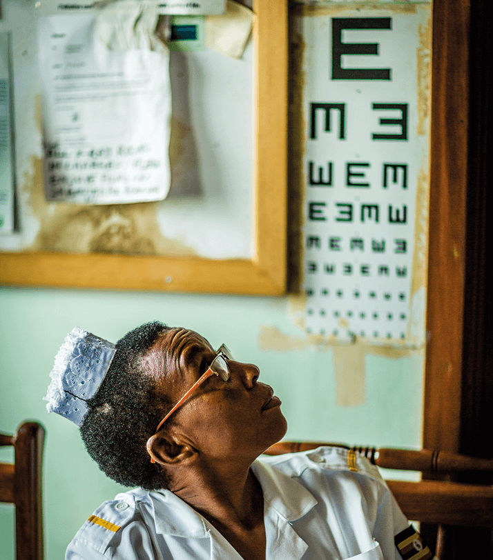
The Vision 2020 Links team – a group of ophthalmologists, orthoptists, and nurses from the Royal Free Hospital in London led by consultant ophthalmologist Clare Davey – went on a 10-day mission to Uganda in April. Photographs documenting that visit are inside this section.

Photographer: Terry Cooper, Volk Optical.

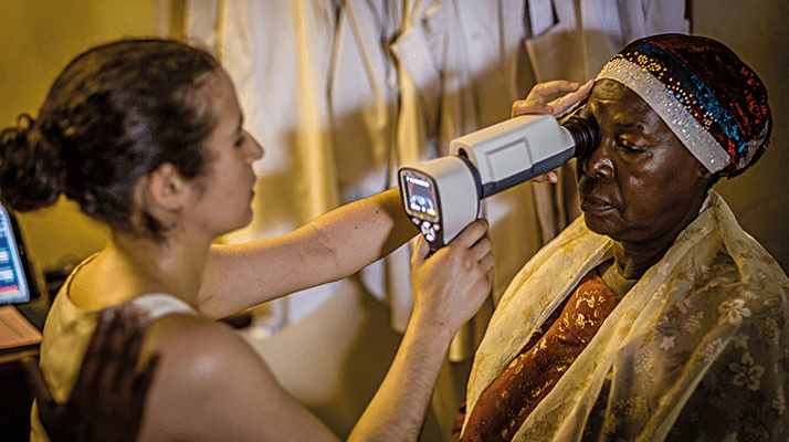
Checking a diabetic patient for eye disease at Mulago Hospital, Kampala, Uganda.

Laser treatment of a patient with diabetic eye disease.

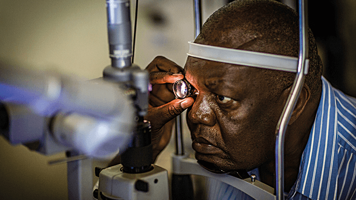
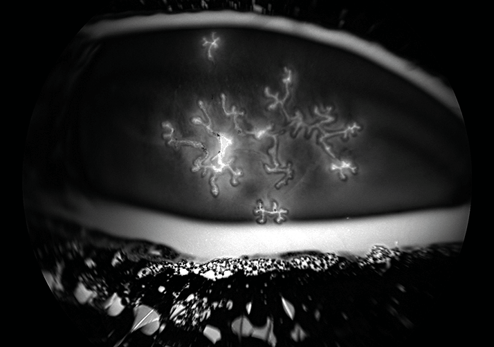
Image credit: Zachary Dupureur & David A Johnson

Autofluorescence of bilateral herpetic dendritic keratitis, dyed using fluorescein strips.

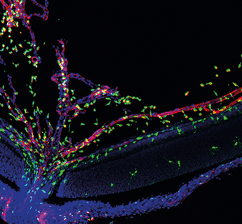
Wai Wong, Chief of the Unit on Neuron-Glia Interactions in Retinal Disease, National Eye Institute, National Institutes of Health.

a. Microglia (green) migrating into the developing retina at postnatal day 3 via the developing retinal vasculature and hyaloid vessels (red).
b. Microglia (green) migrating into the developing retina at postnatal day 7.
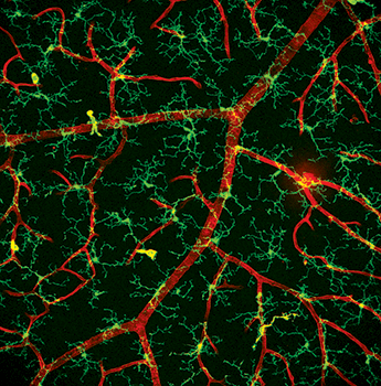
c. Microglia cells (green) in an en-face view of the inner plexiform layer in close proximity to retinal capillaries (red).
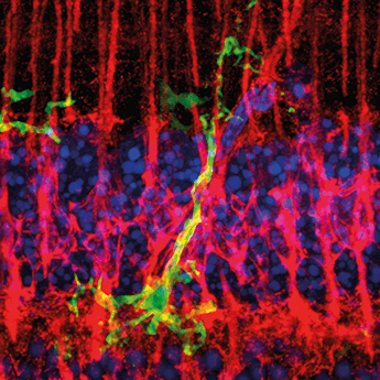
d. Microglial cell (green) fasciculating closely with radial Müller cell processes (red) in an endotoxin model of retinal inflammation.

a
b
c
d

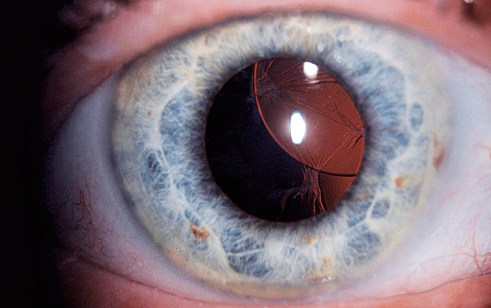
Photographer: Bohdan Kousal, Department of Ophthalmology, Charles University, Prague, Czech Republic

A superiorly subluxated intraocular lens.

Images of Ophthalmology
Images of the Posterior Segment

About the Author(s)

Mark Hillen

I spent seven years as a medical writer, writing primary and review manuscripts, congress presentations and marketing materials for numerous – and mostly German – pharmaceutical companies. Prior to my adventures in medical communications, I was a Wellcome Trust PhD student at the University of Edinburgh.

More Articles by Mark Hillen

Roisin McGuigan

I have an extensive academic background in the life sciences, having studied forensic biology and human medical genetics in my time at Strathclyde and Glasgow Universities. My research, data presentation and bioinformatics skills plus my ‘wet lab’ experience have been a superb grounding for my role as a deputy editor at Texere Publishing. The job allows me to utilize my hard-learned academic skills and experience in my current position within an exciting and contemporary publishing company.

More Articles by Roisin McGuigan

Michael Schubert

While obtaining degrees in biology from the University of Alberta and biochemistry from Penn State College of Medicine, I worked as a freelance science and medical writer. I was able to hone my skills in research, presentation and scientific writing by assembling grants and journal articles, speaking at international conferences, and consulting on topics ranging from medical education to comic book science. As much as I’ve enjoyed designing new bacteria and plausible superheroes, though, I’m more pleased than ever to be at Texere, using my writing and editing skills to create great content for a professional audience.

More Articles by Michael Schubert

Related Content

Newsletters

Receive the latest Ophthalmology news, personalities, education, and career development – weekly to your inbox.

Newsletter Signup Image

False

Advertisement

False

Advertisement

Explore More in Ophthalmology

Dive deeper into the world of Ophthalmology. Explore the latest articles, case studies, expert insights, and groundbreaking research.

False

Advertisement
The Ophthalmologist
Subscribe

About

  • About Us
  • Work at Conexiant Europe
  • Terms and Conditions
  • Privacy Policy
  • Advertise With Us
  • Contact Us

Copyright © 2025 Texere Publishing Limited (trading as Conexiant), with registered number 08113419 whose registered office is at Booths No. 1, Booths Park, Chelford Road, Knutsford, England, WA16 8GS.

Disclaimer

The Ophthalmologist website is intended solely for the eyes of healthcare professionals. Please confirm below: