
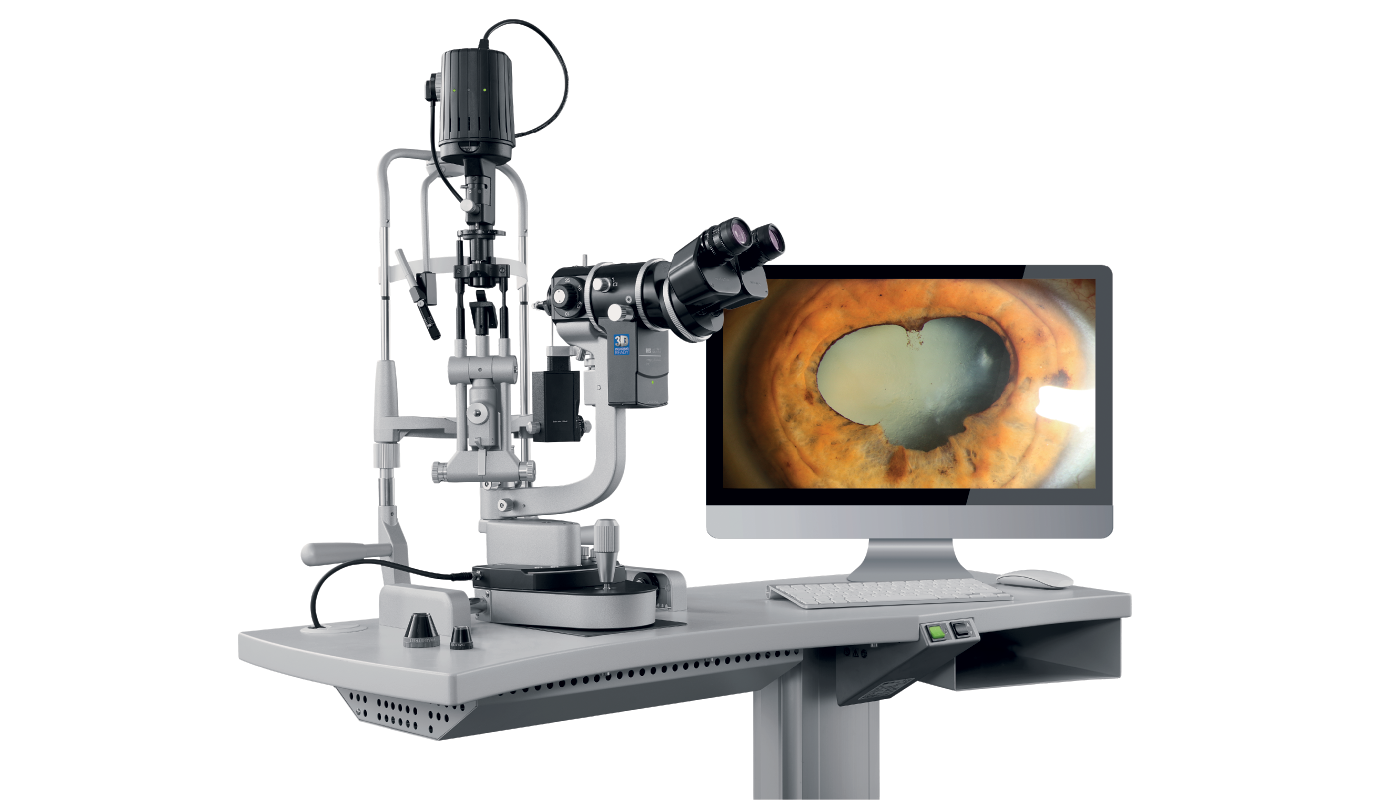
In 2022, Haag-Streit launched the Imaging Module 910 solution for the BQ 900 slit lamp, boasting smart features such as auto-exposure mode and automatic aperture control to improve the success rate of conclusive images offered by conventional slit lamp cameras. In May this year, Haag-Streit further enhanced the proven benefits of the IM 910 by adding a 3D Imaging Option. By enabling the recording of videos and images in 3D, clinicians can now experience a more detailed and authentic representation of the slit lamp exam.
Two eye care experts currently using the 3D functionality of the Imaging Module 910 3D (IM 910 3D) are Michael E. Snyder, MD, of the Cincinnati Eye Institute, Ohio, USA, and Sunil Mamtora, FRCOphth, of Bristol Eye Hospital, UK. Snyder has a private practice specializing in complex cataract, cornea, and anterior segment surgery. Mamtora’s subspecialty interests include cataract and medical retina/uveitis; his practice includes working in the hospital’s emergency department, treating patients with “a huge spectrum of diagnoses and pathology.”
A self-described early adopter of the technology, Dr. Mamtora has been using the IM 910 3D for around 18 months. He considers himself “a slit-lamp imaging enthusiast.” While his hospital has a dedicated imaging team (and a Haag-Streit BX 900 slit lamp, commonly known as “The photographer’s slit lamp,”) Mamtora enjoys taking his own photos and videos.
“The IM 900 was able to capture brilliant photographs,” he says, “but I felt that the process of capturing a photo that I was happy with often took too long – this is a problem in a very busy hospital!” With the latest imaging module, the IM 910, however, “capturing a perfect image doesn’t add any time to my workflow at all,” Mamtora continues. “I can be examining a patient and think, ‘It’d be useful to take a photograph.’ Then all I need to do is turn a knob to enable the camera and then press the capture button. Everything is automatic; there are no settings to change, no PC is required, there’s no waiting for software to load, and the picture looks perfect.”
Smart image capture enables clinics to implement a better digital workflow and viewing images away from the slit lamp supports improved ergonomics
Another benefit is “the seamless way in which I can show patients photographs of their own eyes,” Mamtora adds. “They really appreciate this, and as a result, they understand their condition better, are hopefully more compliant with their treatments, and will have better outcomes.”
Dr. Snyder is a long-time (15-year) user of the IM 900; recently, he has been introduced to the IM 910 3D. He says: “The resolution of the device is high enough and the 3D voxels are good enough to complete the eye exam entirely without oculars for the robust majority of cases. Individual circulating anterior chamber white blood cells or red blood cells (RBCs) can be readily visualized and RBCs can be seen on the 3D screen as they flow through perilimbal capillaries.” This level of digital resolution, without being coupled to the oculars “significantly improves the ergonomics of the slit lamp exam,” he observes. “The high-quality digital record holds the promise of capturing the entire clinical exam remotely, which could eventually improve efficiency for physicians and reduce waiting times for patients.”
The smart choice
The IM 910’s smart features – including an algorithm that auto-selects the best image for the user – brings the technology one-step closer to “an entirely automatically captured slit lamp imaging protocol,” says Snyder. “The auto-selection algorithm seems like a ripe opportunity to take advantage of AI. If we can fully digitize the slit lamp exam, AI diagnostic tools will not be far behind.”
For Mamtora, the intelligent image processing and capturing software are “quite magical.” He explains: “I can look through the oculars without turning away from the patient to look at a screen – when I see something that I think would be clinically useful to photograph, I can just press a single button. In the vast majority of cases, the photos and videos that are captured are exactly what I wanted – without the need to change any settings or distract myself from my examination.”
He adds: “The IM 910’s smart features have changed the way many of my colleagues view slit lamp imaging – from a chore to something that is relatively effortless.”
Documentation: “A picture speaks a thousand words”
The IM 910’s addition of 3D vs “flat” images enriches the documentation process while enhancing the quality of materials. “The presence of 3D gives you more confidence to look at a photo and video and question what more could be added by examining the patient directly,” explains Mamtora. “The most beneficial aspects of 3D are being able to ask for advice from senior or specialist colleagues who haven’t examined the patient themselves and to show patients their own examination in a more immersive way.”
Snyder adds that ophthalmologists are “used to visualizing anatomy in 3D. This system brings that microscopic anatomy within the grasp of patients and their loved ones and, for the first time, allows the surgeon to complete an examination in an entirely digital modality.”
The IM 910 3D can also be integrated into many different environments, such as Haag-Streit’s EyeSuite, or ophthalmologists’ EMR or DICOM systems – providing, says Snyder, “an excellent way to document complex pathology for future comparison and, of course, become a pre-requisite for the remote digital exam.” With the device also permitting documentation of the state of the eye at the time of exam, he adds, it ultimately “fulfills the old axiom that ‘a picture speaks a thousand words.’”
“The other benefit,” says Mamtora, “is for patients to see their own slit lamp examination. They love this, and it’s rewarding for the ophthalmologist. I recently saw a patient who had been referred with a suspicious iris lesion. I was able to take a video and then instantly show the patient the lesion on a 3D tablet.”
Teaching and learning: “Aha” moments
One of the reasons behind Mamtora’s passion for slit lamp imaging is its educational potential. In the past, however, he found slit lamp examination “a challenging skill to learn, and I’m sure I’m not alone.” He goes on, “Developing skills in slit lamp examination is not just about being able to operate the slit lamp, but also to recognize what you are seeing. While the ability to record videos of pathology allows learners to see what they are meant to see, this can often take a long time to learn.”
The ability to show patients a 3D image or video of their condition can significantly enhance patient understanding & compliance with treatment
And the addition of 3D “transforms medical education,” Mamtora says. “Students can put on a pair of 3D glasses and watch the examination live in 3D. This has been valuable for training courses we have delivered in the hospital, as well as giving a high-quality learning experience to medical students, optometrists, nurses, and many others! I wish I could have had something like this when I was beginning my career.”
Snyder has previously used monocular “teaching” viewers attached to a beam splitter, but these optical tubes, he says, “are an ergonomic nightmare, and when the examiner moves the slit lamp abruptly, it can bump the eye of the observer. With the IM 910 3D, the examiner and observer are seeing the exact same thing at the exact same time, and without any parallax error. With that, “teaching becomes increasingly effective, whether the examiner is the learner or the observer,” says Snyder. Consequently, he anticipates “a much higher density of ‘aha’ moments.”
The addition of 3D negates the need for “teaching tubes,” making learning slit lamp examination skills more comfortable for students & teachers alike
The IM 910 3D works very well for one-on-one teaching, but it can also extend the benefits to a much larger group of observing learners, limited only by the access to the 3D screen or availability of 3D projection, Snyder continues. “A whole room full of people can simultaneously see the expert clinician’s exam, and with the recording function, the teaching can continue asynchronously without the presence of the patient. Grand Round programs that include a patient exam will no longer need to inconvenience the subject patients.”
The inclusion of 3D imaging allows all the observers to see the same thing, with the same illumination, resolution, and depth, at the same time
The future: teleophthalmology
In recent years, especially following COVID-19, telemedicine/teleophthalmology has been on the rise. One way that Mamtora sees slit lamp imaging, and in particular 3D imaging, supporting teleophthalmology in UK, for example, is in the referral of patients from optometrists to the hospital eye service. “Being able to more effectively triage referrals by realistically reviewing what was seen by the referring optometrist can allow us to manage patients with advice and guidance, rather than bringing patients for hospital appointments that they might not need,” he explains.
Looking beyond nations and borders, using the IM 910 has enabled Mamtora and his colleagues to stream slit lamp examinations globally as part of an online education program. “This technology can let you get an expert assessment of a patient from anywhere in the world.”
For Snyder, working in the US, where the IM 910 3D is already approved for use, teleophthalmology is a reality, rather than a future aspiration. “Remote consultation, whether local or distant, will likely usher in an environment in which the physician may first meet a patient in person in the operating room.” This increased efficiency will become a welcome modality to deliver needed care on a timely basis. Snyder goes on, “Remote, long-distance consultation may prime the patient and physician for an eventual in-person visit.” What’s more, it can bring “extreme subspecialty care to patients who may not be able to travel to tertiary referral centers either due to cost, time, health, or even visa or immigration limitations.”
From improving access to specialist care in the developing world to strengthening the efficiency of patient pathways within healthcare systems, the potential for slit lamp imaging technology to transform ways of working is significant, agrees Mamtora. He concludes, “3D slit lamp imaging from the IM 910 3D is going to provide many new ways of working that we haven’t even thought of yet!”
The capture of high-quality, high-resolution 3D video images allows consultants to perform teleconsulting in different locations and time zones without limitation
