Janin Lou C. Billano (Headhsot supplied by author)
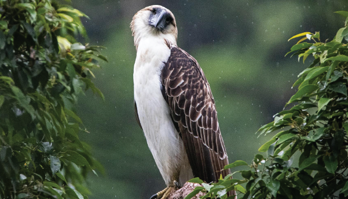
After a serene afternoon filled with the fragrant scent of mountain soil drenched in rain, the night gently covered the shady trees of the bird sanctuary. My mind was filled with fascination and anticipation. That evening, I was lucky enough to be joining a team of ophthalmologists tasked with examining a majestic Philippine Eagle.
The eagle, named Lipadas, was rescued in Davao City, Philippines, by the Philippine Eagle Foundation (PEF). He was first identified in October 2022 when a tribe leader reported that a young Philippine Eagle had crashed to the forest floor along the Lipadas river. The eagle had sustained an injury to his right eye. For the next 12 months, he was observed to make frequent and aggressive “food begging” calls, indicating he was still reliant on his parents for food. By then he was two years old and expected to be fully independent, but his eye injury had made hunting difficult.
A team of biologists, veterinarians, and animal keepers from PEF and the local community was deployed to rescue Lipadas in January 2024. He was safely caught and brought to a local veterinary clinic. Bayani Vandenbroeck, a PEF veterinary consultant, then reached out to our team at the Southern Philippines Medical Center Department of Ophthalmology to perform an ocular examination on the eagle. At present, there are no vets specializing in ophthalmology in our local community.
“Eagle-eyed” is an expression that denotes a sense of perceptiveness to fine, intricate details. An eagle’s vision is exceptionally sharp, allowing them to see small details from extreme distances. They also have two foveae – a deep, central one, and another shallow temporal fovea. In addition, the fovea has one million cells per mm,2 as compared to 200,000 per mm2 in humans. This helps for enhanced visual acuity and higher resolution of sight. Eagles’ eyes are as large as humans and take up almost half of their skulls. They have excellent 20/5 vision, essential for spotting prey from the sky a few kilometers away. Their eye sockets are strategically positioned in the cranial vault to be 30 degrees from the midline of the face. This allows a 340-degree visual field, helping them to achieve excellent peripheral and binocular vision. Eagles who have eyesight defects cannot hunt effectively, and hence, starve to death.
That night, together we were truly in awe as we got up close to Lipadas. We started the examination by instilling anesthetic drops to the right eye and subsequently placing a lid speculum to facilitate adequate visualization of the orbit. There was an obvious loss of orbital volume. Grossly, the injured eye was filled with dried mucus discharge. After cleaning the periorbital and external eye area, we were able to do a portable slit-lamp examination. The right globe was already phthisical. A part of the crystalline lens was also seen sticking out of a big scleral laceration near the limbus. The right eye was already lost. The left eye, however, was perfect.
The injury was clearly traumatic, but the exact cause could not be determined. Philippine Eagles in the wild are threatened daily by human activities. We feared that the injury might have been caused by someone shooting an improvised gun, although no pellets/bullets were seen lodged anywhere in the eagle’s body.
Because of his injury, Lipadas cannot be safely released to the wild. He will remain in captivity and join the PEF breeding program. His circumstances reflect the harsh threats for eagles in the Philippines. Conservation efforts are aimed to alleviate habitat loss, forest degradation, and wildlife hunting. As of now, there are only about 400 pairs of Philippine Eagles left in the wild.
I felt sad as I realized the magnitude of this man-made tragedy. But towards the end of the examination, as I stared once again into the eagle’s ocean blue-grey eye – Philippine Eagles are the only raptors in the world with bluish-grey eyes – I knew that the rescue was a major accomplishment. Lipadas’ expressive gaze was like a comforting hug after an exhausting day.

Credit: Rowell Taraya
Acknowledgments
We would like to thank the Philippine Eagle Foundation, especially Jayson Ibañez (Director of Operations), Bayani Vandenbroek, and Nielsen Donato (veterinary consultants), Dominic Tadena (Senior Animal Keeper), and the biologists and staff who made the rescue and rehabilitation of Lipadas a success. Finally, we thank the communications team of PEF and Keith Bacongco for the photos used in this article.
Sources
DENR, 2019. Updated National List of Threatened Philippine Fauna and their Categories. Quezon City: DENR Administrative Order.
Philippine Eagle Foundation, 2009. Raptor Research and Management Techniques with a Focus on the Philippine Eagle. Davao City: PEF.
“Raptor Ophthalmology: Anatomy of the Avian Eye,” LafeberVet, 26 Oct. 2020, lafeber.com/vet/raptor-ophthalmology-anatomy-of-the-avian-eye/.
