Though ophthalmology is an exciting field full of innovation and advancements, many of these developments are founded on long-lasting technologies and principles. This is particularly true for retinal imaging. Most imaging modalities used today in multimodal devices involve the incorporation of principles that have been used separately for years with the aim of seeing more, more clearly. With a technique capable of pseudo-three-dimensional (3D) visualization of retinal images, NIDEK has developed a novel, unique, and clinically beneficial imaging modality, Retro mode. The innovation uses a concept previously undeveloped in the retinal industry, demonstrating a new way of pushing forward to meet an enduring goal.
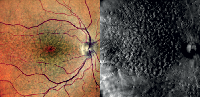
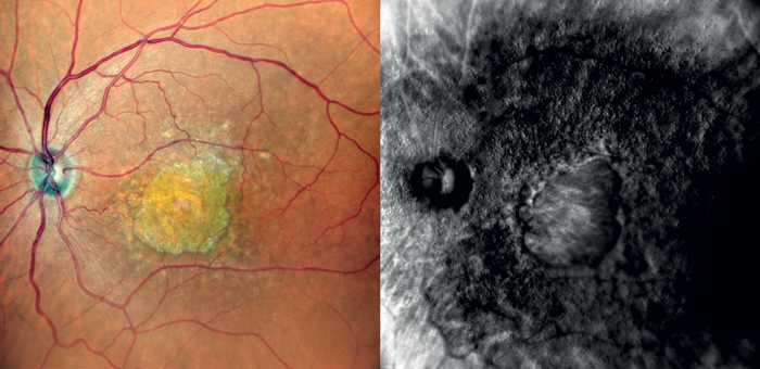
Retro mode is one of the imaging modalities incorporated into the Mirante Scanning Laser Ophthalmoscope (NIDEK). This imaging modality uses a near-infrared wavelength (790 nm) and a laterally deviated confocal aperture with a central stop for obtaining pseudo-3D images of deeper retinal layers. It detects pathologic changes in the choroid caused by drusen, edema, and other subtle chorioretinal pathologies. This wavelength is particularly suited to imaging chorioretinal pathologies because of its deeper penetration within the retinal layers. Alongside this, the confocal apertures, which can be placed circularly or laterally shifted to either the left or right, enable the collection of backscattered light which is then converted to pseudo-3D images. Together, these imaging principles allow excellent image contrast and result in better visualization of the depth and breadth of pathology compared to commonly used retinal imaging devices, such as optical coherence tomography (OCT) or color fundus photography.
So, what does this mean in practice? Retro mode can be applied in many indications in the immediate short, medium, and long term. With an aging population there is increasing prevalence of – and public health concern regarding – AMD; early detection and timely management and treatment of AMD, and its hallmark of drusen, is thus needed more than ever. The unparalleled ability of Retro mode to detect subtle drusen – as well as better delineate the appearance of a variety of drusen types and the area of the retina affected by pathology makes it a crucial retinal visualization tool for combating increased AMD prevalence. Additionally, the recent approvals of treatments for geographic atrophy have resulted in an increased demand for effective ways of monitoring the efficacy of treatment – a demand that Retro mode can meet.
With the Classification of Atrophy Meetings program recommending multimodal imaging as ideal for assessing patients with atrophy resulting from AMD, it is clear that Retro mode is particularly well-suited as a complementary imaging technology to be included in multimodal imaging for diagnosis, monitoring and management of both AMD and geographic atrophy.
As mentioned earlier, innovation is a continual process, something NIDEK is acutely aware of which is why they continue to actively engage with a working group of specialists who use Retro mode. Through incorporating key feedback from these practitioners, NIDEK continues to not only enhance the imaging and diagnostic capabilities of this innovation, but also shape the future through the dreams and aspirations of the past.