Surgical Tips for Retina – Part 2
In the second of this two-part series, Ferenc Kuhn and Andrzej Grzybowski discuss what the ophthalmologist needs to address during eye surgery
Ferenc Kuhn, Andrzej Grzybowski | | 5 min read | Discussion
Credit: Shutterstock.com
Retinal surgery has gradually become vitreoretinal (VR) surgery; it’s extremely rare today that a posterior segment surgeon performs scleral buckling only. This two-part series, therefore, discusses a handful of highly selected surgical tips related to vitreoretinal procedures. In Part 1, we covered the preparation needed before surgery begins.
No procedure, especially VR surgery, should be performed without the surgeon first designing a plan. The plan is akin to a map – how to get from place A to place B. The surgeon should have a rough idea of the eye’s condition prior to surgery, and the anatomy to arrive at as the surgery is completed. This strategy mostly concerns preparing for what is in the “road” that connects the two endpoints. Naturally, the plan is always modified based on how the actual surgery evolves and how tissues react to the surgeon’s manipulations.
Here, we review several tips that are useful during surgery.
1. Placement of the (intrascleral) cannulas
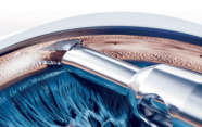
The role of the three cannulas is mainly to provide access to the vitreous cavity for the infusion line and for the instruments to be used. The trocar at the time of scleral penetration should have a ~15-degree angle to the surface; its distance from the limbus in an emmetropic eye is 3–3.5 mm, and they are ideally positioned in the right eye at 7 o’clock (infusion line; 5 o’clock in the left) and at 2:30 and 9:30 for the “working” cannulas. A fourth cannula is inserted, typically superiorly, if truly bimanual vitrectomy is performed.
2. Anterior segment: the lens
When the strategy has been formulated, the surgeon must take into consideration that every VR surgery leads to cataract development (not only “if the patient is over 50 years old,” as the literature used to claim). In other words, cataract development is not a complication but a side effect of vitrectomy. In patients compromised by age, concomitant cataract surgery should be offered, especially if silicone oil is to be used. The old counter-argument that cataract surgery increases the severity of the postoperative inflammation is no longer valid with today’s phacoemulsification technology.
3. The vitreous: the sequence and completeness of the removal
If there is no severe vitreoretinal traction, and especially if the vitreous is transparent (for example, no vitreous hemorrhage is present), a posterior-anterior approach is recommended. The vitreous removal is started centrally and posteriorly, and is gradually extended towards the mid-vitreous cavity and then towards the periphery and the retrolenticular space. For this latter area, vitreous removal is highly advantageous if there is severe VR traction (proliferative vitreoretinopathy [PVR], proliferative diabetic retinopathy [PDR]) and in eyes with retinal detachment or trauma. In a phakic eye, small air bubbles should be injected behind the lens; if they remain there, vitreous is still present behind the lens.This is first detached by gentle aspiration and then removed by holding the vitrectomy probe’s port sideways, minimizing the risk of lens touch/cut.
4. Posterior vitreous detachment (PVD)
One of the most misleading concepts in the literature has been the myth of spontaneous PVD developing in conditions such as vitreous hemorrhage, retinal detachment, PVR, PDR, and trauma involving the posterior segment. However, if the surgeon actively tries to verify the presence of vitreous on the posterior retina – and triamcinolone is an almost mandatory vehicle for this – they will discover more often than not that what appears to be PVD is in fact vitreoschisis (a thin layer of vitreous is still undetached from the retinal surface). If this is not detached and removed, a host of complications (from macular pucker to retinal detachment) may arise.
The most common method of surgical PVD-creation involves using high flow/aspiration but no cutting, as the vitrectomy probe is dragged along the disc margin on the temporal side, barely touching or just above the retinal plane. It is triamcinolone that helps the surgeon visualize the vitreous, both when it is still attached and when it finally pops up. Once separation has been achieved, the cutting action is to be activated so that no VR traction is transmitted towards the periphery where separation of the two tissues is impossible.
If aspiration is insufficient to lift the posterior vitreous, a barbed needle/blade can be used to engage the vitreous; this is ideally done under a contact lens to increase the resolution and avoid retinal damage.
5. Macular pucker
A forceps or a barbed needle/blade can be used to lift the scar tissue from the retinal surface – almost all eyes with a pucker have a true PVD. Care must be taken if there is an area in the scar that seems to be very prominently elevated, especially if it is whitish colored – this may be a retinal fold. The entire membrane should be detached within the vascular arcade, and serious consideration should be given to also remove the internal limiting membrane (ILM). The ILM may already be partially broken, and, more importantly, its removal prevents recurrence of the pucker (otherwise occurring in up to 10 percent of the eyes).
6. Surgery on an injured eye
Surgery on an injured eye is always a challenge because of the many unknowns and the relatively small number of rules related to management. Moreover, this surgery might require different experience or training based on different cases. Below we touch upon two very basic issues of the tissue in the tactics category.
- Anterior segment: hyphema
If liquid, the blood can be removed through a single paracentesis created on the temporal side. The cannula is used both to push down the opening’s inferior lip and to irrigate the anterior chamber (AC). If the blood is clotted, an AC maintainer must be positioned first, ideally inferotemporally and with its tip slightly downwards over the iris (to prevent damaging the endothelium and the lens). The vitrectomy probe is used to engage and remove the clot; the port should be kept sideways and always occluded by the clot.
- Anterior segment: the iris
This diaphragm must always be reconstituted if damaged; should posterior segment surgery be required, the reconstruction of the iris is the last step, only when the VR situation is deemed final. If during the initial examination/surgery a wide pupil is found, the iris may be missing (rupture) or simply retracted by fibrin (contusion). In the latter case the iris must urgently be pulled using serrated vitrectomy forceps so that the fibrin “glue” is broken and a normal-sized pupil created.
Ferenc Kuhn is Professor of Ophthalmology at the University of Alabama, Director of Clinical Research at the Hellen Keller Foundation, and Head of the Department of Ophthalmology, University of Pécs Medical School, Hungary.
Andrzej Grzybowski is a professor of ophthalmology at the University of Warmia and Mazury, Olsztyn, Poland, and the Head of Institute for Research in Ophthalmology at the Foundation for Ophthalmology Development, Poznan, Poland. He is EVER President, Treasurer of the European Academy of Ophthalmology, and a member of the Academia Europea. He is a member of the International AI in Ophthalmology Society (https://iaisoc.com/) and has written a book on the subject that can be found here: link.springer.com/book/10.1007/978-3-030-78601-4.
