The Sooner, the Better
Why early screening is essential in the fight against keratoconus
Identifying keratoconus early is our biggest challenge, as changes in corneal shape typically occur before there is a loss of best-corrected vision. As well, the slit lamp exam is typically normal in mild keratoconus as well as in many cases with even moderate keratoconus. Since loss of best-corrected vision is often a later consequence, patients with keratoconus can often have the condition for a significant period of time or experience significant changes in corneal shape before they are diagnosed. There are a few reasons for this: first, it takes a while for patients to recognize their sight has changed, and even longer for them to decide to see an ophthalmologist or optometrist.
Since keratoconus can be asymmetric, some patients can see and function normally with their less affected eye despite the contralateral eye experiencing significant loss of vision. Second, when patients with early keratoconus who still have good vision see an eye care professional, they often only undergo a standard eye exam, where early signs of keratoconus are easily missed. As a result, many patients with keratoconus are actually only diagnosed at eligibility screenings for refractive or cataract surgery when a screening topography is performed. Unexpectedly, these exams have become a useful means of catching keratoconus in a mild or earlier stage, before the condition becomes too advanced.
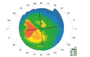
The issue of keratoconus diagnosis is shared by optometrists and ophthalmologists alike. The fact is that we do not offer routine topography screenings to every patient who enters our clinics, but only in cases when there is vision loss or irregularity of the cornea. And maybe we should. My colleague’s 19-year-old son recently developed increased astigmatism in one of his eyes. His eye doctor was wise enough to know that, when patient’s experience a significant change in astigmatism, you should perform a topography screening. And it was fortunate he did; the doctor diagnosed very early stage keratoconus. As with all eye conditions, knowing when – and who – to test is critical.
Early intervention is crucial; the sooner we catch the condition, the sooner we can perform cross-linking to stabilize the cornea and prevent progression. Though we can cross-link patients with moderate to advanced keratoconus who have already experienced vision loss, they may be forced to wear special contact lenses or glasses. And that’s why clinicians are trying to catch people before their best-corrected vision drops to 20/40 or 20/60 – we want to diagnose them when they are still 20/20. If we want our patients to lead a normal life, we need to be responsive to any subtle suggestion that keratoconus may be present. If a patient’s experience a significant shift in astigmatism power or axis, screen them with topography.
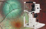
Unfortunately, a lack of affordable equipment is holding us back. Not every eye care location has topography available – and even those with the right technology may not offer it to every patient. My hope is that industry helps create affordable devices, so that patients and eye care providers everywhere can have access to screening tests. I’m currently working with a company to develop an inexpensive topography unit that will hopefully cost around a thousand dollars with a small monthly cost for cloud-based integrated AI – affordable for even the smallest practice.

Cataract, corneal and refractive specialist at the Center for Excellence in Eye Care in Miami, Florida, USA.
