The 2014 Innovation Awards Are here

Ophthalmology is one of the most intense incubators of medical innovation. Competition is truly driving innovation, and this is resulting in not just the incremental improvements in products that you might expect, but also some big, game-changing leaps too.
Here we recognize a year’s worth of innovation. Apps, IOLs, vitreous cutters, imagers and lasers – the latest and greatest of these are all there.
But which one came out on top?

1. iolAMD
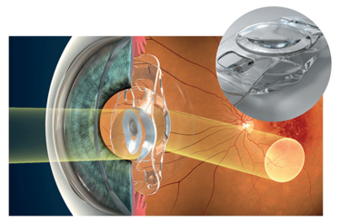
The world’s first micro-incision, injectable telescopic implant
Produced by: London Eye Hospital Pharma (www.iolamd.com)
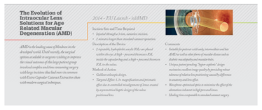
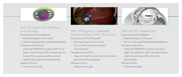
Detail: The iolAMD procedure involves two advanced hydrophobic acrylic IOLs being injected into the eye using modern surgical techniques via a 3 mm, sutureless incision. The two lenses work together and act like a Galilean telescope, gently magnifying the image entering the eye and diverting it to a healthier part of the retina. The section of healthier retina then takes over the role of the macula and provides the iolAMD patient with significantly improved vision. The magnification achieved is around 1.3×, which allows for bilateral implantation, and while visual acuity is improved, visual field is maintained.
The iolAMD lenses contain patented hyper-aspheric surfaces and unique wavefront characteristics that reduce the optical distortions that are normally associated with high powered lenses, as well as creating an increased tolerance of relative lens positioning. This additional positional tolerance maintains image quality, even if the lenses end up slightly closer or further apart due to the physiological variances of each individual eye.
Impact: iolAMD is a new IOL system for the treatment of early, intermediate and end-stage dry AMD and other macular pathologies including diabetic maculopathy, macular holes, myopic degeneration and hereditary retinal diseases such as Stargardt’s and Best’s.
AMD is the leading cause of blindness in the developed world and there is currently no cure, but iolAMD can restore central vision in this large group of potential patients, greatly improving their quality of life.
Invented by leading eye surgeon Bobby Qureshi and developed by optical physicist r Pablo Artal, iolAMD represents a huge leap forward in patient safety and postoperative optical performance. This procedure is as safe as routine cataract surgery and can be used as an exciting new alternative to monofocal lens implants in appropriate patients with macular disease.
Judges’ comments: “This technology could allow blind individuals to see better.”
“Big benefit for patients with AMD. Combines two principles of magnification and paramacular image.”
2. Cassini Total Corneal Astigmatism
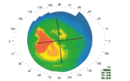
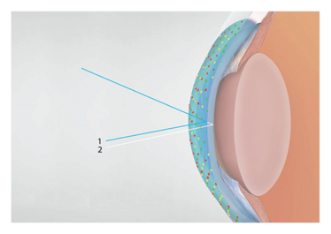
Measures the total corneal astigmatism – anterior and posterior – for optimal selection and alignment of premium IOLs
Produced by: i-Optics (www.i-optics.com)
Detail: Failing to take into account posterior corneal astigmatism (PCA) during cataract surgery may lead to incorrect estimation of total corneal astigmatism (TCA). Research has shown that selecting toric IOLs based on anterior corneal measurements could lead to over-correction in eyes that have with-the-rule astigmatism and under-correction in eyes that have against-the-rule astigmatism – but there seems to be a large variety in the relationship between anterior and posterior astigmatism.
What this means is that patients undergoing cataract surgery would benefit from individual measurements of the TCA (anterior and posterior) rather than using a generic nomogram. Cassini provides the personalized data that enables the ophthalmologist to create unique, personalized surgical plans for each and every patient.
Impact: A study by Warren Hill demonstrated that over 50 percent of patients with cataract patients have anterior corneal astigmatism that falls within the range correctable by toric IOL – but nomogram estimates of PCA can lead to intra or post-operative refractive surprises. Taking into account the individualized measurements of TCA means that physicians are better able to select the most appropriate lens and axis for their patient, and provide more confidence and better outcomes – which should ultimately lead to a higher volume of premium patients coming to your clinic.
Judges’ comments: “Improves refractive outcomes. Period.”
3. ViaOpta Nav
An app to help blind and low vision people with their mobility
Produced by: Novartis International AG (www.novartis.com)
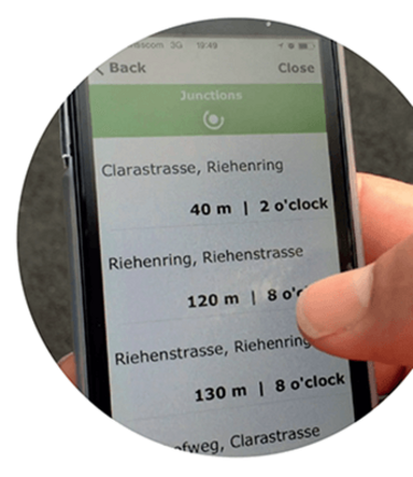
Detail: The aim of the app is to allow blind or low vision individuals to move independently, with the ability to walk to a destination and have information useful to facilitating their orientation while they are moving. The user can enter a destination and get turn-by-turn directions, and waypoints can be added to improve the effectiveness of the calculated route. While moving, the app will also give information on junctions as well as distances and directions. The user can query the app at any time for their position and get it in terms of street address. A list of junctions around the user, with the corresponding distances and bearings can also be obtained. The information is provided by text to speech or if it’s running, the screen reader.
Impact: Commercial navigators are of considerable value to people with low vision, but there are a number of features that are either unavailable or inaccessible to these users. ViaOpta Nav picks up the slack. For example, pedestrians with low vision often need to know their position in order to avoid getting lost, to feel safe and be aware. They also find it extremely useful knowing information about the next junction, even if it is that they only need to cross it, without having to take a turn.
The app (available on iOS and Android systems) gives this information using an extremely easy and immediate user interface for a person with vision loss – and could be an indispensable to thousands of visually impaired people in their daily mobility tasks.
Judges’ comments: “Important public health impact.”
“Great opportunity for visually impaired people.”
4. Icare HOME
A tonometer for 24 hour IOP self-monitoring
Produced by: Icare (www.icaretonometer.com)
Detail: Designed for home use in patients with (or clinical suspicion of) glaucoma, who need regular 24-hour IOP monitoring per their ophthalmologist’s recommendation. The device is based on a rebound measuring principle that requires no topical anesthesia, air or specialized skills. It includes a number of features that make it easy to use: automatic measuring sequence, intelligent positioning and automatic OD/OS recognition display for ease of use. The results are not displayed to the patient and can only be retrieved by a healthcare professional using Icare LINK software.
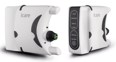
Impact: One of the first methods for 24 hour IOP monitoring outside the clinic, Icare HOME enables IOP self-monitoring anywhere and at any time. The concept of continuous monitoring should provide more information for the ophthalmologist – and comfort for the patient. A better understanding of the patients’ IOP profile may also enable the ophthalmologist to further optimize their patients’ medication regimens, enabling improved IOP control and clinical outcomes.
Judges’ comments: “Great innovation for IOP self-monitoring”
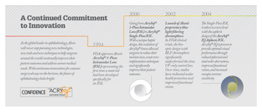
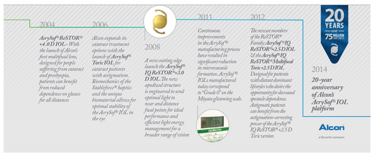
Timelines
5. UNO Colorline MACH2 Vitreous Cutter
A double bladed vitreous cutter for fast core vitrectomy with no traction and safe shaving
Produced by: Geuder AG (www.geuder.com)
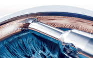
Detail: The MACH2 double-blade vitreous cutter improves can considerably improve the performance of vitrectomy. The guillotine blade carries out two cuts per work step, meaning that compared to single-blade vitreous cutters, it slices the vitreous into smaller pieces – improving not only vitrectomy performance, but aspiration performance and flow rate too – and has the happy side-effect of increased blade durability. Furthermore, even when you’re cutting near the periphery of the retina, the blade performance is so good, the retina remains virtually completely immobile.
You also get a smoother cut – when using a single blade the surgeon must control two independent parameters, vacuum/flow and cut rate, and the higher the cut rate, the lower the aspiration flow. With a single blade, the aspiration window is, cumulatively, closed for longer – but with the MACH2, the aspiration window remains permanently open and decouples cut rate from aspiration flow, resulting in faster core vitrectomy and fully controllable vitreous shaving.

Impact: The constant, high flow, along with a permanently open cutting window, an any-time adjustable flow, and double blades that allow for up to 12,000 cuts per minute helps the surgeon perform a fast and safe core vitrectomy with minimal traction at the vitreous base – and makes duty-cycle management obsolete. The pulse-free action also results in predictable behavior of the retina and therefore increased patient safety. The MACH2 also provides optimal complication management in complex indications such as trauma, organized vitreous, vitreous hemorrhage or luxated lenses.
Judges’ comments: “Sounds like an obvious thing to do – but a significant advance.”
6. LipiView II
The only device available which assesses both meibomian gland function and structure
Produced by: TearScience (www.tearscience.com)
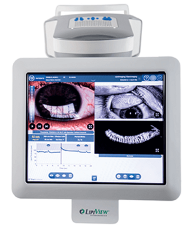
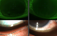
Detail: LipiView II assists clinicians by improving the diagnosis of meibomian gland dysfunction (MGD) through the provision of an unparalleled objective examination of patients’ tear film lipid layer thickness, blink profile, and dynamic meibomian imaging (DMI). DMI simultaneously employs dynamic surface illumination and adaptive transillumination, eliminating glare and providing auto-adjustment for lid thickness. Dual mode imaging delivers a more accurate visualization of the meibomian gland structure – allowing physicians to evaluate gland structure, measure lipid layer thickness and determine partial blinking with confidence.
Impact: To properly diagnose and educate patients, eyecare providers must examine gland structure and function with as much accuracy as possible. LipiView’s complete, detailed gland images (made possible by their proprietary illumination technology) also helps convey to patients the importance of treating the glands before the disease progresses. Around 90 percent of all dry eye can be attributed to MGD, and with 300 million people suffering from dry eye worldwide, MGD may represent one of the largest patient segments that could present to an eyecare practice. LipiView II’s sophisticated visualization helps clinicians identify root etiology in patients with dry eye, take proper management steps and improve their quality of life.
Judges’ comments: “Novel and interesting”
7. SP.eye
Intravitreal injection assistant device, combining control and sharps safety
Produced by: Salar Surgical (www.salarsurgical.co.uk)
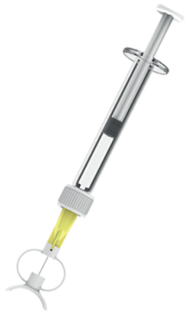
Detail: SP.eye provides three-dimensional control of needle position with respect to the limbus, depth and angle of injection, and is the first device to feature integrated sharps safety, with both passive needle tip protection and active locking. It is supplied mounted on a standard 30 G needle, and can immediately be integrated into existing workflow patterns.
Impact: SP.eye increases safety for both patient and clinician, and allows nurses and other non-surgeons to deliver repeatable injections with confidence. This complements the shift towards nurse injectors, and could increase capacity and efficiency in medical retina clinics; fitting directly into existing workflow patterns. It can be used with both ranibizumab and aflibercept, and is compatible with push fit, Luer lock and proprietary pre-loaded syringe systems. It is also the first sharps safe intravitreal injection device, and therefore the only one to comply with European regulations on reduction of needle stick injuries.
Judges’ comments: “A clever and useful device.”
8. IC-8 small aperture IOL
An IOL that provides extended depth of focus by exploiting the small aperture principle
Produced by: AcuFocus (www.acufocus.com)
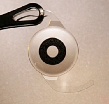
Detail: A single-piece hydrophobic-acrylic IOL with an embedded mask measuring 3.23 mm in total diameter, with a central aperture of 1.36 mm. The mask contains 3,200 micro perforations to minimize diffraction effects. The IOL extends depth of focus through the use of the small aperture principle – the mask only allows the central paraxial light rays to reach the retina and restricts the defocused light that reduces image quality. The IOL is implanted monocularly, and the fellow eye can remain phakic if the crystalline lens is clear, or be implanted with a high quality monofocal if lens opacity is present.
Impact: When a patient undergoes cataract surgery they are typically treated with a monofocal IOL – limiting their ability to see clearly at distance only. To view objects up close these patients require reading glasses. This is why presbyopia-correcting IOLs were developed – to address this need. However, today’s multifocal and accommodating lenses come with significant limitations such as photic phenomena, incomplete range of vision or unpredictable functionality. The small aperture is a proven method for improving range and provides patients with continuous functional vision from near-to-far. Glare and halos complaints are minimized as there aren’t competing focal points (as there are with multifocal IOLs), meaning that this IOL has the potential to change the way we treat patients with cataract and provide a reliable vision correction method.
Judges’ comments: “Simple and Novel”
9. KXL II System
Topography-guided custom cross-linking for refractive applications
Produced by: Avedro (www.avedro.com)
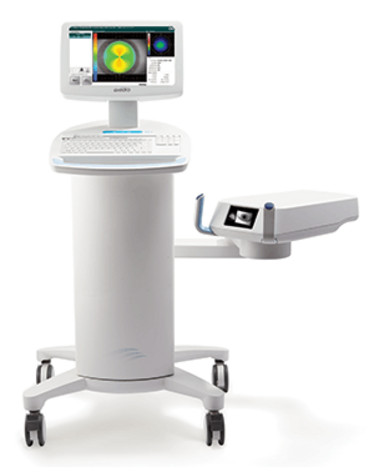
Detail: By leveraging the fundamentals of traditional cross-linking for keratoconus, Avedro developed a new application – the use of accelerated cross-linking alone as a refractive treatment. The process, photorefractive intrastromal CXL (PiXL), is performed using Avedro’s KXL II system. The treatment requires no cutting or contact – it just involves the activation of riboflavin eye drops with ultraviolet light.
Corneal topography is integrated with real-time eye tracking and programmable UV-A illumination patterns, allowing treatment that is customized for each individual patient. CE marked, and in clinical use since April 2014, PiXL has now been performed on over 150 patients across 17 centers, with encouraging early results. The technique has the potential to treat post-cataract ametropia, provide non-surgical refractive correction, and maintain or improve corneal biomechanical integrity.
Impact: PiXL is the first procedure to offer non-surgical refractive correction with cross-linking alone, thus eliminating the corneal weakening that is inherent in all traditional approaches to refractive surgery – and has the potential to be the most significant development in refractive correction since LASIK surgery. PiXL could potentially be used for two of the largest applications in ophthalmology: post-cataract ametropia and primary refractive correction – which represent 10 million procedures each year, and have a combined yearly revenue potential of $2 billion. There are over 90 million patients in the US with myopic error of -2.4 D or less, 99 percent of whom do not have laser refractive surgery. With PiXL, refractive practices have the potential to offer those patients a non-surgical solution to their vision correction needs, which could truly be revolutionary.
10. Tecnis Symfony Extended Range of Vision IOL
A presbyopia-correcting IOL which corrects by extending range of vision rather than using traditional multifocal technology
Produced by: Abbott Medical Optics (www.abbottmedicaloptics.com)
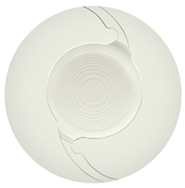
Detail: The first and currently only lens in a new range of presbyopia correcting IOLs (PC-IOLs) that extends the range of vision to correct presbyopia. The unique and proprietary design combines diffractive echelette and achromatic designs, elongating the range of defocus while correcting chromatic aberration to improve image quality. This results in truly continuous vision that could provide independence from spectacles for most tasks. Importantly, the extended range of vision is accomplished with an incidence of glare and halo comparable to a monofocal IOL. In a recent study, patients achieved visual acuity of 20/20 or better across 1.5 D of defocus and 20/40 or better across a 2.5 D range of defocus.
Impact: The Tecnis Symfony offers cataract patients with presbyopia an opportunity to achieve spectacle independence after surgery and employs technology that can provide quality vision at all distances. With strong visual performance and a low incidence of dysphotopsias, it may increase the appeal of surgical presbyopia correction – globally, an estimated 15.4 million patients with cataract are candidates for PC-IOLs, but only around 4.5 percent are expected to receive them. Because it has strong visual performance and a low incidence of dysphotopsias, the Tecnis Symfony IOL may increase the appeal of surgical presbyopia correction, thereby allowing more cataract patients to benefit from the advantages of advanced, PC-IOL technology.
11. WIOL-CF: Bioanalogic IOL
A bioanalogic polyfocal IOL for correction of cataract and presbyopia

Produced by: Medicem (www.medicem.com)
Detail: Building on the heritage of Otto Wichterle, the inventor of hydrogel contact lenses, Medicem has leveraged over 20 years of scientific and clinical research into its proprietary WIGEL hydrogel material that has been specially developed for intraocular applications, creating the WIOL-CF, the first bioanalogic polyfocal IOL for correction of cataract and presbyopia. By mimicking the natural crystalline lens in material, size and design, WIOL-CF is designed to deliver visual quality at all distances. Smooth hyperbolic aspheric optics, with no multifocal refractive or diffractive zones, in combination with biocompatible material allows the patient to perceive a natural transition of vision, while maintaining contrast sensitivity and long-term functionality.
Impact: The market for presbyopia correcting (PC) IOLs has grown dramatically, more than doubling between 2008 and 2012, but PC-IOLs still only represent around 3 percent of total IOLs implanted globally. Many surgeons believe this is due to performance limitations and negative trade-offs such as optical phenomena and low contrast sensitivity, restricting the candidate patients for PC-IOL implantation to only very highly motivated patients who wish to be spectacle-free. Bioanalogic WIOL-CF offers an appealing solution to these problems and holds the potential to substantially grow the PC-IOL market.
Judges’ comments: “A new concept of polyfocal IOL to create spectacle independence.”
12. Orion
Device-independent OCT image analysis software
Produced by: Voxeleron (www.voxeleron.com)
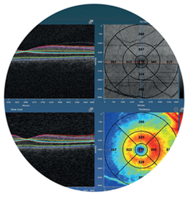
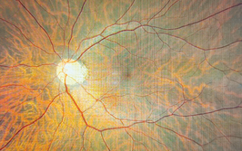
Detail: OCT is the standard of care in ocular disease management, but is underutilized as current software provided by the OCT manufacturers support measurements of three (of several) retinal layers at most. The retina is an extension of the central nervous system and its deeper, neuronal layers have been shown to help gauge not only the health of the eye, but also to offer direct correlates to brain structure and health. These layers are more challenging to segment, and cannot be measured with existing software. Orion addresses this need with device-independent segmentation of seven retinal layers, including the inner and outer nuclear layers, and has been validated by two independent studies. It also provides automation, speed, and intuitive interaction for an optimized workflow.
Impact: Well established in ocular imaging, OCT is now poised to become an important tool in the fight against neurodegenerative diseases including ALS and Alzheimer’s. OCT is likely to become a ubiquitous, front line disease screening and management tool impacting millions of people, but only once analysis software can support it. The technology within Orion is an important step in this direction, that should help accelerate the pace of discovery in ophthalmology and neuroscience, and empower clinical researchers to study the relationship between the neuronal layers of the retina and a wide variety of neuropathies.
Judges’ comments: “Good alternative in hospitals with different OCT manufacturers to have same software for evaluation.”
13. OPMI LUMERA 700 and RESCAN 700
Providing ophthalmic surgeons ZEISS HD-OCT images of the eye without interrupting surgery
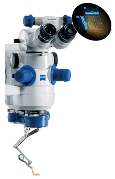
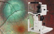
The RESCAN 700 has a broad range of applications in anterior and posterior segment surgery.ZEISS HD-OCT images add a real-time third dimension to the visualization capabilities directly in the eyepiece of the OPMI LUMERA 700 surgical microscope. It provides surgeons with unprecedented views below the surface of the surgical field, enabling them to see more – even transparent structures – and helping them back up their decisions and improve their surgical technique, without compromising surgical workflow.
By merging two gold standards into one system, ZEISS has created a first – a surgical microscope with integrated intra-operative OCT: the OPMI LUMERA 700 and RESCAN 700.
Impact: Today’s surgeons may have difficulty seeing certain anatomic details during surgery. With this new visualization tool, ophthalmologists can overcome these limits. Surgeons now can see even transparent ocular structures during surgery, monitor progress during a procedure, and verify clinical results in the OR. OCT scans can also be stored and recalled for later review and “fly through” via CALLISTO eye from ZEISS. Simply put, the new device enables better decision-making during surgery.
Judges’ comments: “Enables direct control of both anterior and posterior segment procedures.”
14. OCULUS BIOM ready
The world’s first single-use wide angle viewing system
Produced by: Oculus Surgical (www.oculussurgical.com)
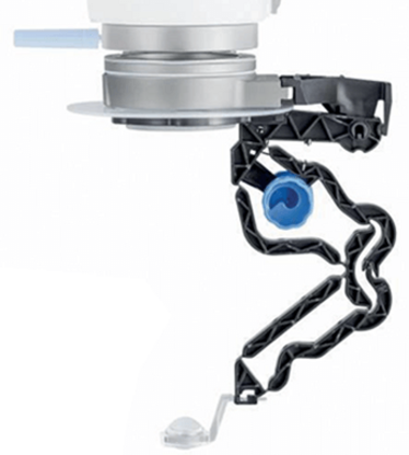
Detail: Provides the perfect view for non-contact wide angle observation for retinal surgeons without using a contact lens. Easily connected to the microscope; while the surgeon is observing the vitreous and the fundus, the BIOM ready is aligned coaxially with the operating microscope… but during extraocular surgery phases, it’s swung out of the observation beam. It incorporates the new BIOM HD Disposable Lens for unparalleled visual clarity and provides excellent depth of field for better stereopsis. The OCULUS BIOM ready comes pre-assembled with the BIOM HD Disposable Lens in a sterile blister pack.
Impact: Provides the optimal balance between efficiency and high optical quality, with outstanding resolution in the periphery, often reducing indentation during laser. The depth of field is increased over other wide angle systems, allowing the surgeon to perform macula work without the need for a contact lens, saving time and money. Sterilization “down time” is decreased – increasing OR efficiency – and as it’s a single-use device, it reduces the risk of cross-contamination.
Judges’ comments: “Amazing wide field view in a disposable lens.” “Improves convenience.”
15. Navilas 577+
Tissue-friendly, standardizable, navigated microsecond pulsing therapy for retinal disease
Produced by: OD-OS (www.od-os.com)
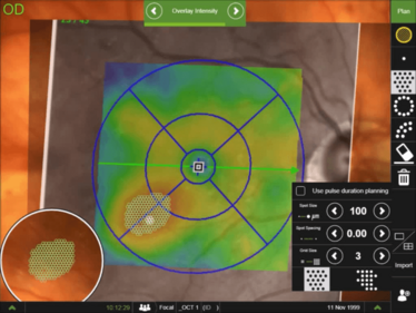
Detail: Laser energy is split up into a pulse train of low-energy pulses that stimulate the retina, but do not heat the tissue to a coagulation threshold – and as a consequence, retinal function loss and scarring can be avoided. Navilas is the only navigated microsecond pulsing therapy (NMPT) system that allows navigated application of this advanced subthreshold laser technique; the treatment area can be precisely delineated based on imported OCT thickness maps, and the aiming beam is prepositioned – compensating for eye movement and allowing complete coverage without undefined overlap.
The treatment is documented in real time, providing visual feedback about treated areas and degree of completion. An initial case series at LMU Munich performed by Marcus Kernt showed no tissue damage or retinal function loss (as expected) with this method. Contact lens-free application and comfortable infrared illumination make this a patient-friendly therapy and set Navilas 577+ apart from slit lamp-based lasers.
Impact: Retinal laser is making a comeback in diabetic eye disease because of the chronic use and expense associated with anti-VEGF therapy. Initial studies indicate that laser treatment can add durability to anti-VEGF gains and reduce patient burden. The technique further refines retinal laser therapy: inter-operator variability is minimized by OCT-based planning with real-time documentation, and retinal tissue function is preserved by microsecond pulsing. NMPT has the potential to become the standard adjunct to anti-VEGF in diabetic eye disease.
Judges’ comments: “Great laser to improve patients’ outcomes.”
I spent seven years as a medical writer, writing primary and review manuscripts, congress presentations and marketing materials for numerous – and mostly German – pharmaceutical companies. Prior to my adventures in medical communications, I was a Wellcome Trust PhD student at the University of Edinburgh.
